Cultivating cells in conventional two-dimensional (2D) systems to mimic in vivo situations is a challenge. In this environment, not only cell-to-cell communication is lost, but also mechanical and biochemical properties. The goal of 3D (three-dimensional) cell culture is to provide an environment that accurately resembles the complex habitat that surrounding cells experience in their native tissues, allowing for in vitro growth, differentiation and functionalization.
3D platforms consisting of natural polymeric nanofibers are being increasingly used in areas such as tissue engineering, regenerative medicine, stem cell research and oncology, not only due to their structural similarity to the Extracellular Matrix (ECM), but also due to their high batch-to-batch consistency, their ability to shape biomaterials into different geometric shapes and the absence of animal derivatives.
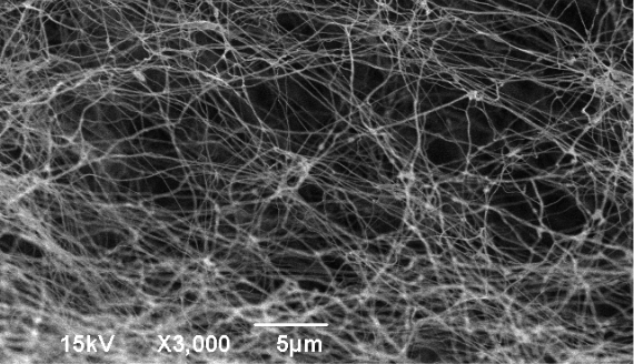
Human collagen - Electron micrography
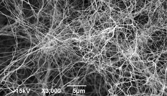
Nanofibers of the 3D CellFate® matrix
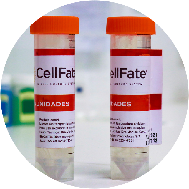
The 3D CellFate® matrices developed by Biocelltis are biomaterials made up by natural polymeric nanofibers from 50 to 100 nm in diameter, similar to human collagen, biocompatible, sterile and ready-for-use, ideal for growing human and animal cells in research labs. Biocelltis offers 3D CellFate® matrices for a wide range of shapes and sizes of culture plates for routine use in laboratories, in addition to customized solutions.

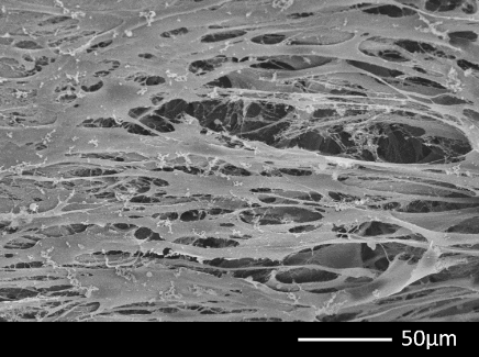
L929 fibroblasts cultured on the 3D CellFate® matrix. Image obtained using scanning electron microscopy (SEM).
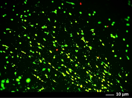
Human umbilical vein endothelial cells (HUVEC) cultured on the 3D CellFate® matrix and stained with the Live/Dead reagent. Green staining shows viable cells (calcein) and red staining shows dead cells.
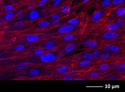
Primary human fibroblasts cultured on the 3D CellFate® matrix with highlight to the cell nucleus stained in blue (DAPI) and cytoskeleton stained in red (phalloidin). Image obtained using confocal microscopy.